Echinoderms are an interesting bunch of critters, encompassing intertidal favorites such as the star fish, the sea cucumber, and the sea urchin. The roots of the word "Echinoderm" stem from the Green word "ekhinos," meaning porcupine or hedgehog, and the root -derm, meaning skin. In my opinion, sea urchins represent the epitome of a spiky-skinned animal, and in light of that fact I thought I'd look at a few spines under the SEM.
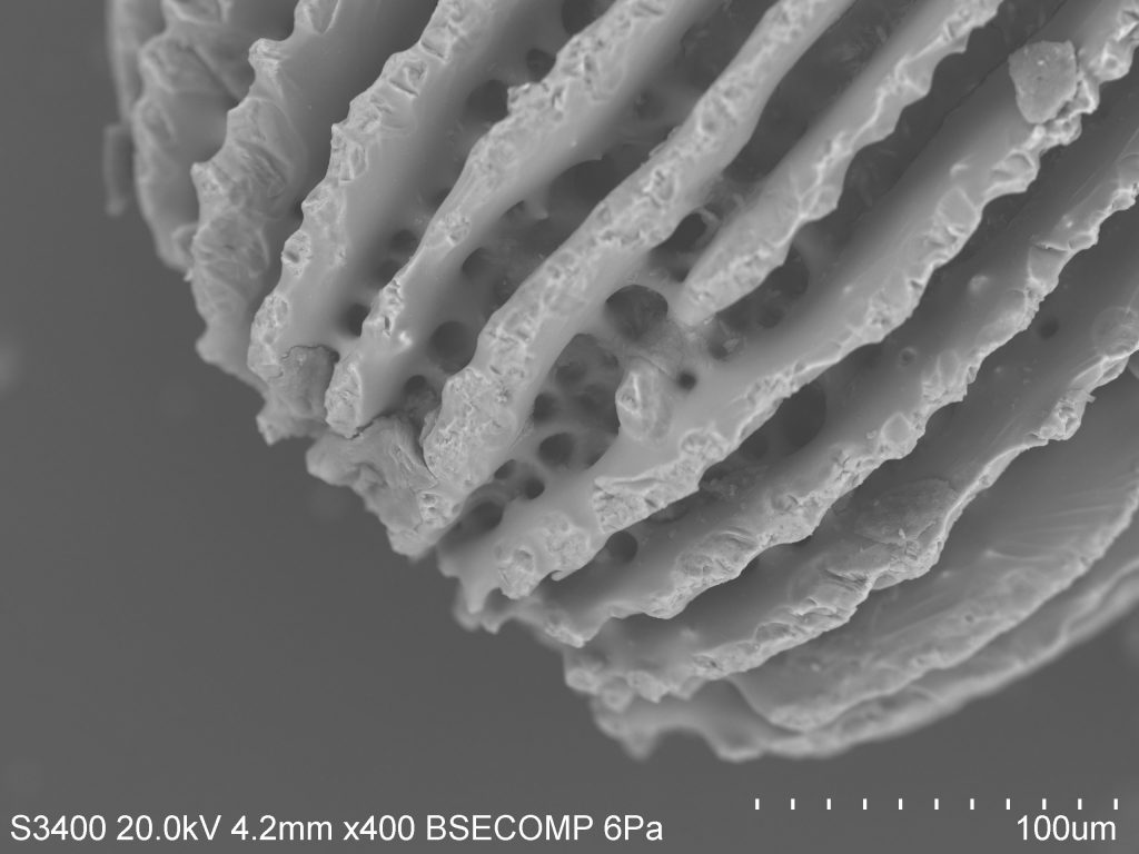
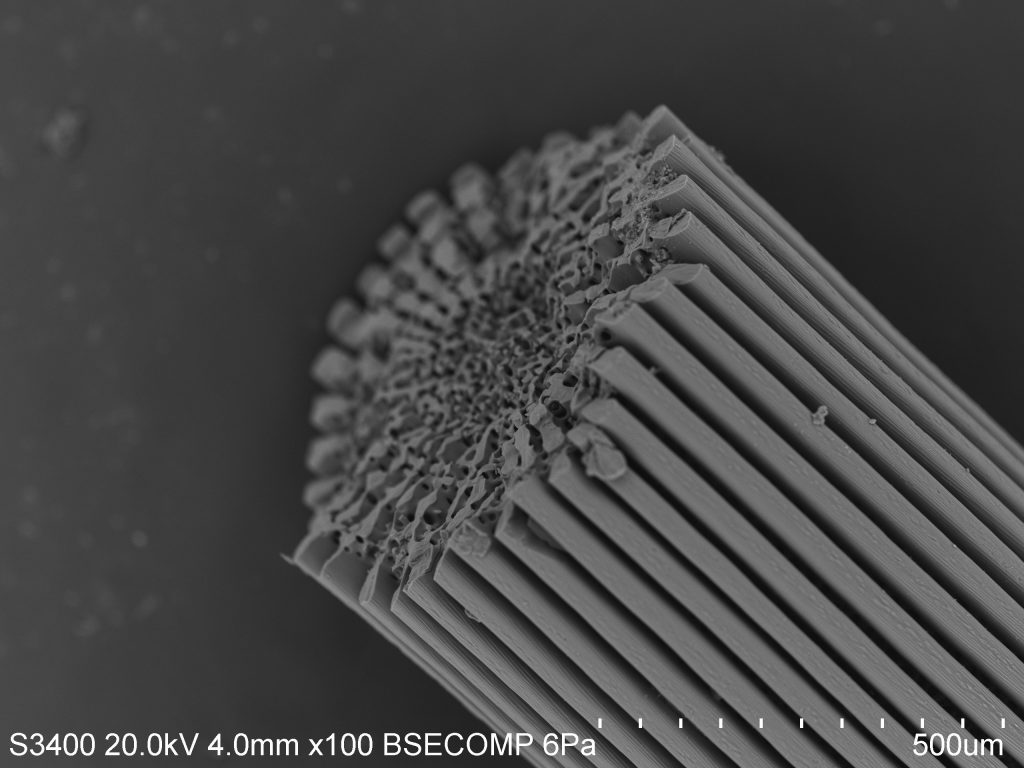
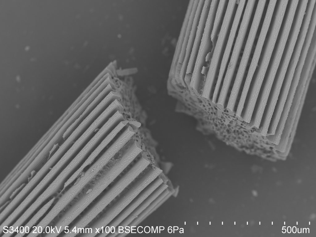
As you can see, the spines are ridged and porous. Sea urchins do not have a closed circulatory system and instead rely on an open water vascular system to help with the uptake of nutrients, the flushing out of wastes, locomotion, and respiration. As such, much of their body plan is open to the environment. In this case, it is actually covered by a thin layer of epidermis (the outer layer of cells), making the spines themselves a feature of the interior endoskeleton. You can think of this as similar to the skin that covers our bones, although spines and bones serve very different purposes.
The name for this internal structural organization is the stereom. The stereom is a feature that all echinoderms share, and is useful in identifying lineages in fossilized samples. Spines mainly show two distinct morphological configurations: the base, made of a meshwork stereom, and the shaft, which has several longitudinal septa and a central core of meshwork stereom. This can be seen distinctly in the photo. The stereom is sponge-like and varies in composition by species. In a sea urchin, the stereom may be 50% living cells such as connective tissue cells and phagocytes involved in nonspecific defense. The rest will be composed of a matrix of calcite sclerocytes which direct mineral production and repair.
Over several days, sea urchins are actually able to regrow spines in places where damage has been inflicted, or to grow entirely new spines. The urchins begin growing micro-spines in a conical shape by precipitating calcite from the surrounding seawater. As these small spines thicken, lateral growth takes place, allowing neighboring micro-spines to join in a horizontal "bridge." The formation of bridges results in the mesh of cells spanning from the base of the spines to the tip.
All this terminology is only the tip of the iceberg (tip of the spine?), and I'm keen to look further. The test of the sea urchin, which is the "hard shell" under the spines and feet and skin, is full of microscopic pores through which several accessory structures pass. The geometry is quite interesting from a macro-perspective, as all echinoderms share pentaradial symmetry, meaning their bodies are grouped loosely into five identical parts. In urchins, these manifest as alternating ambulacral (related to the feet) and inter-ambulacral (between the feet) regions. I imagine the micro-analysis of differences between these regions would be interesting as well.


