Piston Core 26

Previous posts in the New Guinea region have discussed sites adjacent to delta fronts. Nearshore sediments are typically variable in nature consisting of sand, mud, pebble-sized particles, and biogenic material. Here, we will focus on piston core 26 (PC26), a drill site furthest from the shoreline.  PC26 was extracted south of Manus Island at a water depth of 875 m and a subsurface depth of 48-49 cm. Sediment deposition in deep water environments, located on the abyssal plain, build up at reduced rates. These decreased rates of sediment accumulation are due to the limiting factors of particle transport; volcanic ash and windborne particles are slowly transported towards the open ocean environment. Therefore, we should expect sediment extracted from PC26 to be largely homogenous.


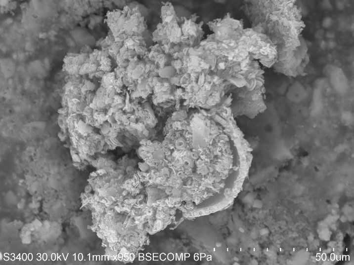
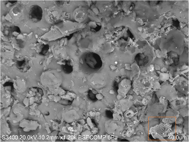
Immediately, it is apparent that sediment from PC26 is predominantly biogenic material. The image above highlights several chambered tests, or shells of foraminifera. Their test are typically cemented with sand grains or other materials, and crystalline CaCO3 in the form of calcite or aragonite depending on the species. This particular species has large pores and exhibits a trochospiral chamber arrangement. A mature foraminifera may range from 100 µm to 200 cm in length. There are approximately 4,000 species of forams, although only a few modern species are in existence, making these microogranisms the most abundant shelled protists in the marine environment since the Cambrian period.


At close inspection, we can see an aggregate of coccolithophores settled within a foraminifera test. There is a distinctive difference in the size and shape of these two microorganisms that have the identical chemical compositions. Therefore, it is suitable to characterize this pelagic sediment as calcareous ooze, given its calcic nature. The remarkably small size of coccolithophores in comparison to forams does not limit its abundance in pelagic sediments. Calcareous oozes cover approximately one-thirds of the Earth’s entire surface, and due to seawater/carbonate interactions, calcareous ooze begins to dissolve below the calcium carbonate lysocline in the water column. Consequently, material below the calcium carbonate compensation depth calcareous ooze completely dissolves.


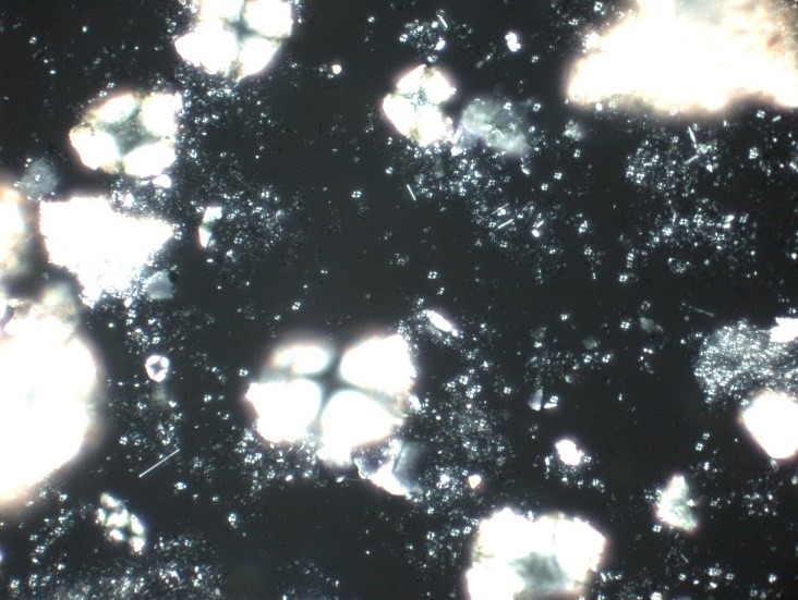
A visual representation of what we see underneath the SEM is also visible in PPL (top) and XPL (bottom). In PPL, the pore-rich tests of the foraminifera are easily distinguishable, particularly in the upper left corner. In XPL their calcite bearing shells illuminate the field of view with their four-chambered anatomy. Similarly, aggregates of coccolithophores are distinguishable in XPL.

Piston Core 36

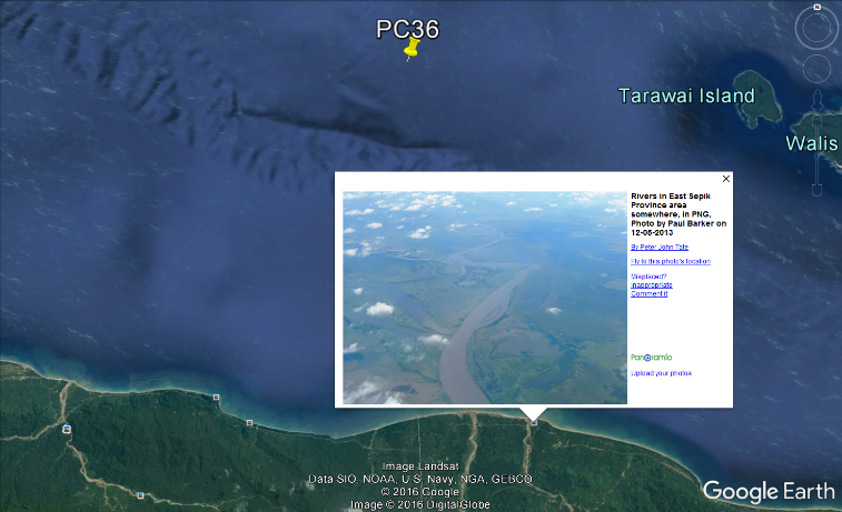
In previous posts we have discussed deep-sea sediments that are rich in skeletal debris. Now we will focus on a site that is abundant in siliciclastic material. Piston core 36 (PC36) is a site located adjacent to the Western Sepik River at a water depth of 903 m. The sample we will be focusing on was collected at a subsurface depth of 139-140 cm. Its close proximity to the delta may the source of such terrigenous material.


The amount of sediment a river can transport changes over time, therefore, sediments found at varying depths can reveal changes in precipitation. Change in precipitation is a result of climate change, and as climate changes so do the environments that are in the region! A comparison of siliciclastic material will be addressed in an upcoming post where we will look at another sample from PC36, but at a subsurface depth of 218-219 cm. Siliciclastic rocks are non-carbonate sedimentary rocks that are almost exclusively silica bearing. They are terrigenous material, derived from erosion or weathering of land-based rocks, transported by wind or water. Common siliciclastic minerals are quartz, feldspars, micas, and heavy element minerals.


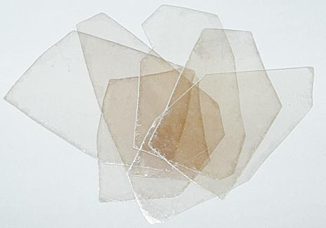
The images above highlight a common sheet silicate mineral, known as a mica. Its platy texture is easily distinguishable on the adjacent minerals in the lower and upper left corners. This platy texture is the result of basal cleavage; minerals in the mica group have one cleavage plane and can be peeled into perfect thin sheets. Mica formations are generally associated with volcanoes and hydrothermal vents. Several of the brighter minerals scattered across the surface of the mica are calcite (CaCO3) crystals.


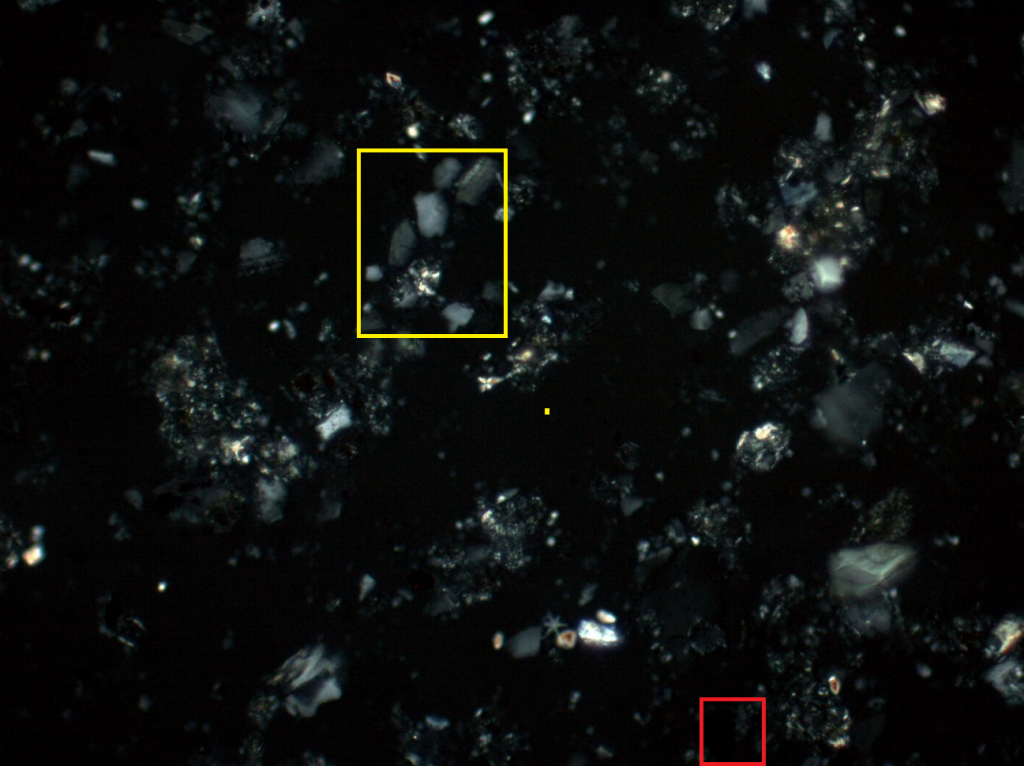
As expected the sediment from PC36 is highly siliciclastic (yellow), but contains very few quartz crystals, in PPL (right) and XPL (left).  Samples containing quartz are very stable and mature sediments. Therefore, this sample appears to be freshly deposited given its immature mineral assemblage. This supports the hypothesis that this sediment may have been transported from land by the Sepik River. This sample also contains a fair amount of opaque (red) minerals. Opaque minerals prevent light from passing through and commonly contain iron oxides and sulpihides. It is apparent that this sediment contains minimal skeletal debris as compared to previous sediments we have looked at. For comparison, see the previous post regarding PC23.


Deep-Sea Minerals

Minerals are found in various shapes and sizes in the deep marine environment. Some minerals form naturally in the ocean through chemical processes, whereas others are transported to the sea from terrestrial environments by wind or water flow. We are going to take a look at a few minerals found at various coring sites in the New Guinea region. With the help of the EDX and knowledge of chemical and physical properties of minerals, at minimum it is possible to determine a minerals group.


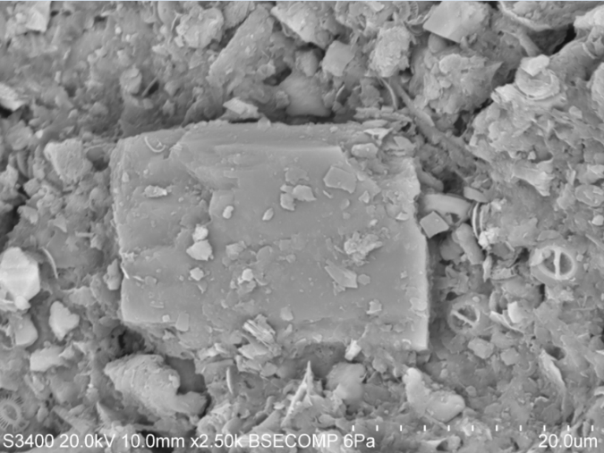
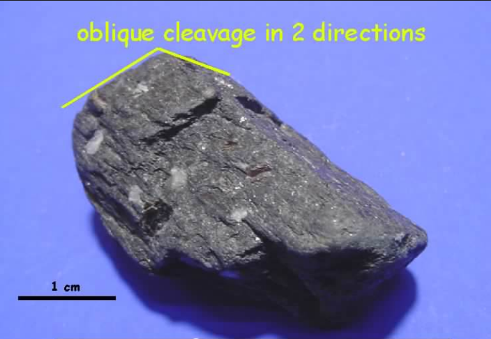
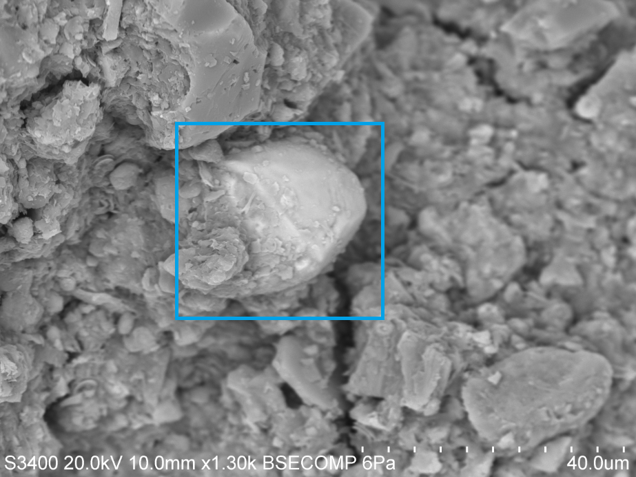
At first glance mineral identification on a SEM seems like an impossible task. However, we can deduce that this mineral likely belongs to the amphibole group. This is largely due to its orthorhombic shape and 120° cleavage plane. In mineralogy, cleavage is used to describe how a crystal breaks on a particular plane. EDX point scan analysis indicates that the mineral contains approximately 3% Al, 7% Fe, 8% Mg, 9% Ca, 24% Si, and 41% O. Since the chemical composition of an amphibole is NaCa2(Mg,Fe,Al)5(Al,Si)8O22(OH)2, we can be certain that this mineral is an amphibole.

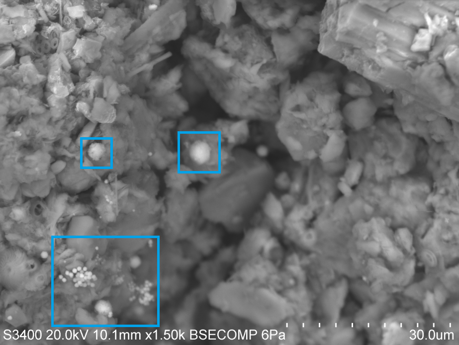
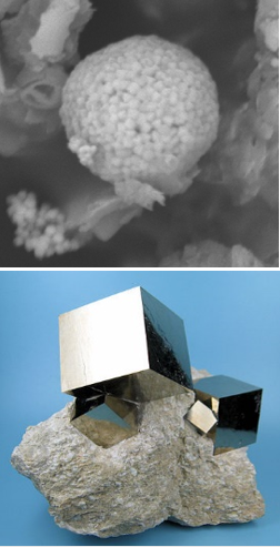
Is that really gold settling on top of marine sediment? Unfortunately, marine sediments are not a lucrative source of gold. What we are looking at here is pyrite (blue), known as fool’s gold. Pyrite has a chemical composition of FeS2 and its formation is largely dependent on sulfate availability in the ocean, where sulfate is converted by bacteria to hydrogen sulfide. Pyrite commonly forms perfect cubes, however in this case they are visualized as spherical aggregates of microcrystals, called framboids.


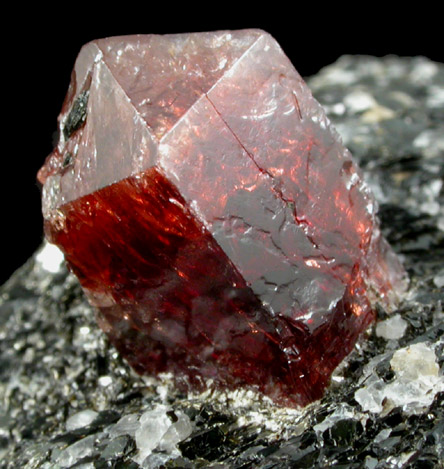
Heavy element minerals are indicators of mature, stable sediments. Since heavy minerals are often found concentrated in or around bedrock material, it is likely that this material is terrigenous. EDX microanalysis shows that this mineral contains approximately 17% Si, 50% Zr, and most of the remainder in oxygen. Based on the analysis, it can be inferred that this mineral displaying conchoidal fracture is a zircon (ZrSiO4) crystal.

Piston Core 23

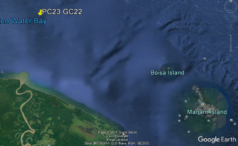
Since we have covered fundamental information regarding microfossil composition and structure, now we can focus on individual core sites and general geology of the New Guinea region. The science crew aboard the Roger Revelle (RR1313) collected a total of 54 cores near the Papua New Guinea margin. Sites PC23 and GC22 are the southeastern most sites, situated east of the Sepik Delta and west of the Manam Volcano. We will be focusing on piston core site 23 (PC23) collected at a water depth of 712 m and a subsurface depth of 431-432 cm.

Tectonically, the New Guinea region is complex due the formation of a subduction zone by rapid (geologically speaking) motion of the sea-floor. Subduction, indicated on the map as black lines with the teeth, is a geological process that occurs when two plate boundaries collide. As the two plates collide the denser slab sinks into the mantle and beneath the lighter slab, where temperature and pressure progressively increase with depth. Under these conditions trapped fluids such as seawater generates magma, which rises into the upper slab, and forms a volcano. In this case, the Indo-Australian Plate is moving northeastward but crashes into the Pacific Plate that is moving west with a slight north component. This vertical and horizontal displacement is geometrically classified as oblique-slip faulting, resulting in the formation of a volcanic chain.


Sediment at PC23 is particularly exciting because it is biologically and lithologically diverse. The image on the right displays several coccolithophores on top of flaky clay-sized particles. The image on the left features several coccolithophores that have situated on top of a “hole bearing” organism, known as foraminifera. A smaller foram fragment (orange) is visible in the lower right. Looking at sediments on a SEM is especially useful for end-member microanalysis. Mineral end-member identification is used to distinguish between biogenic, terrigenous, and hydrothermal sediments, all of  which may be altered when water masses mix and interact with rock.


An EDX detector paired with the SEM is able to generate data in the form of a spectra that displays known elements in the point scan by focusing a high-energy beam of particles into the sample. The output spectroscopy peaks for Spectrum 2 above indicates that calcium is present (peak intensity does not correspond with weight %), verifying that the microfossil analyzed is a coccolithophore.


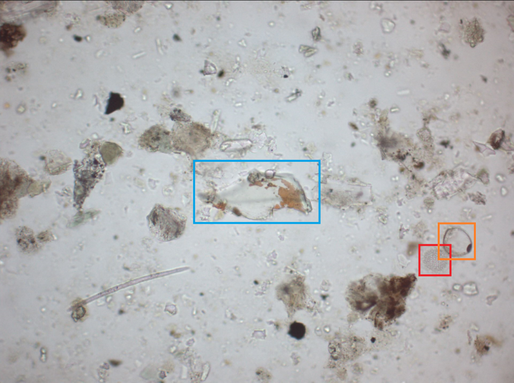
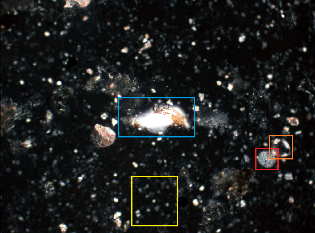
In addition, scientists can analyze sediments underneath a petrographic microscope by preparing smear slides. A smear slide is a thin layer of unconsolidated sediment spread onto a glass slide. Petrographic analysis of a smear slide gives quantitative and limited compositional information with the ability to view the sample in plane-polarized light (PPL, top) and cross-polarized light (XPL, bottom). Under PPL, the sediment from PC23 appears to be clay-rich and contain minimal biogenics. However, under XPL we can see that the sediment is abundant in biogenics relative to the other particles. Coccolithophores (cluster in yellow) are calcite bearing organisms that are only visible in XPL. The dinoflagellate (red) and foraminifera (orange) are visible under both light conditions. The centered shard of vitric material (blue) is an indicator of hydrothermal activity and suggests volcanism. The combined use of a SEM and petrographic microscope make for strong tools to reconstruct ancient oceans.


Biogenic Ooze

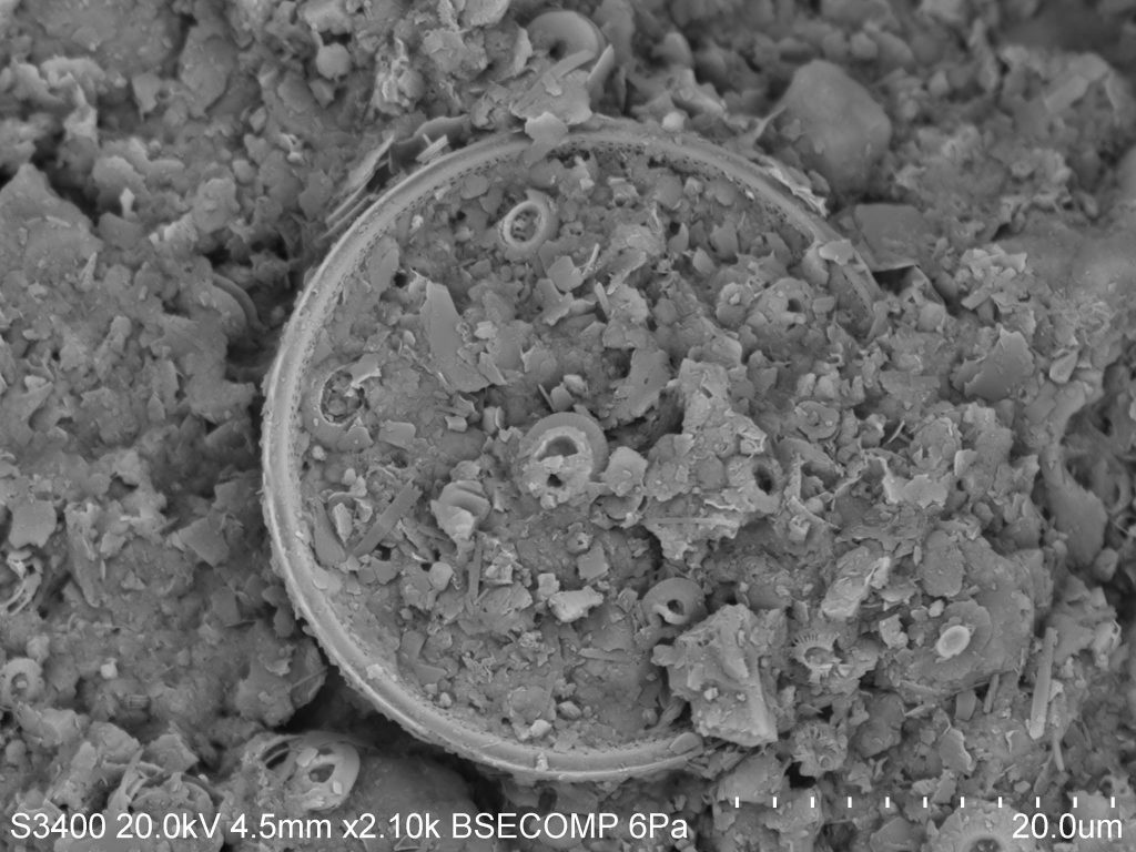
Biogenic sediments extracted from deep-sea sediment cores enable scientists to piece together a story about Earth’s early marine environments and past climate. This particular sample was collected off the coast of Papua New Guinea in the western Pacific Ocean by the International Ocean Discovery Program’s expedition RR1313. Microfossil-rich sediments, such as the one pictured above is termed a biogenic ooze and is characterized by containing greater than 30 percent skeletal debris.


The disk-shaped organism featured above is a well-preserved concentric diatom surrounded by a type of phytoplankton, known as a coccolithophore. In terms of shape and size, coccolithophores are a highly diverse group of phytoplankton that produce calcareous skeletons composed of calcium carbonate (CaCO3) called coccoliths. Whereas diatoms are encased by a hard-shelled silica (SiO2) body, often referred to as opal. The siliceous shells of diatoms allow them to endure turbulent mixing in the upper layers of the ocean. As diatoms bloom and die, they sink to the bottom of the ocean and become exceptionally useful in paleoceanographic research. Microfossils are used to infer rates of sedimentation, biological productivity, nutrient availability and oxygenation events which are imperative to understanding past, present, and future climate.


Foraminifera and Coccolithophores

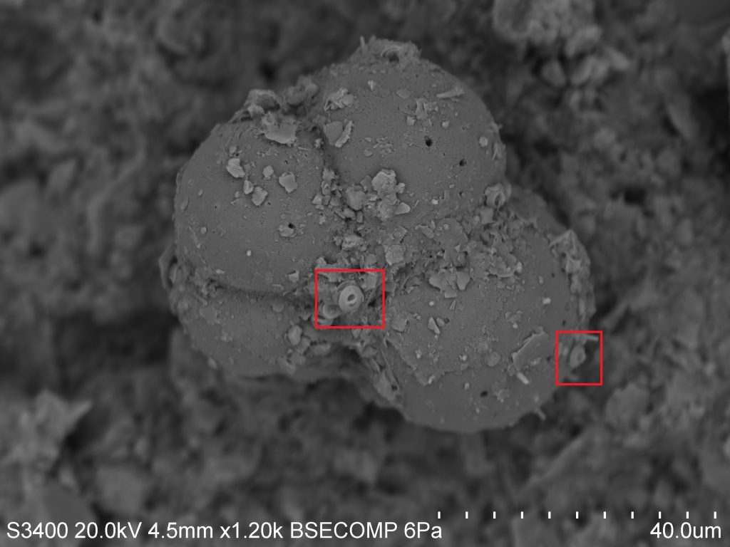
Chances are you'll be seeing a fair bit of discussion regarding plankton as we go forward, because (1) they show up in many of our samples, and (2) there's a lot we can learn by studying them! SEM is especially useful for the study of plankton, as their small size makes traditional compound microscopes somewhat ineffective. This photograph, taken during our first run-through with the SEM, shows an image at 1,200X magnification, as compared to the 100X magnification you might hope to get with a standard scope!


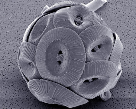
In the above photo we can see examples of both zooplankton (microscopic animals) and phytoplankton (microscopic plants). The first is the obvious, bulbous species in the middle of the image. This is the test, or shell, of a Foraminifera. Forams represent an ancient and speciose group of zooplankton which live mostly in sediment (as is the case here), but also in the water column. Within the red squares you will see a second, smaller phytoplankton species known as a Coccolithophore. The round plates are known individually as placoliths, and bundle together around the algal species to form a coccosphere. I've included the photo below to give you an idea of how they appear when alive in nature. Similarly, coccolithophores inhabit both benthic and open-water habitats.


Plankton species such as these can provide a surprising amount of information not only about themselves, but also about the environment in which they grow.  The tests you see here are made of calcium carbonate (CaCO3) which the organisms "pull" out of the water and use to build their hard parts. As such, isotopic analysis of these tests can tell you about the atmospheric and oceanic conditions at the time of their formation, since the two systems are linked in the carbon cycle. When the organism dies, calcium carbonate sinks to the bottom of the ocean in the form of coccoliths and other tests and become a part of the sediment record, which can be studied later by scientists as part of a sediment core.

There are many other applications in the study of plankton, some of which we will likely cover in future installations. Stay tuned, and remember: It's the little things.