SEM Theory

SEM Theory

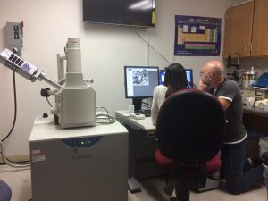
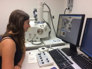
The following is a description of SEM history and function provided by Dr. Ivano Aiello, excerpted from Moss Landing Marine Laboratory's 50th anniversary blog. The full article can be accessed here, and we plan to add an even more thorough discussion in the near future.


To study the small world of the oceans, classic tools of marine science are not enough to observe and collect valid scientific data. The observation of the microscopic features of marine organisms such as corals, foraminifera, diatoms or sponges or the interior structures of organic cells (nucleus, mitochondria…etc.) requires very high magnifications, 10,000 and larger, more powerful than the optical microscopes, limited by the physics of light can yield.


The invention of the first electron microscope by Max Knoll and Ernst Ruska and the production of the earliest scanning-transmission electron microscope (SEM) by Manfred Von Ardenne in Berlin in the 1930s allowed scientists to finally observe the microscopic world to magnifications before unthinkable. The introduction of the first commercial scanning electron microscopes (SEMs) in 1965 opened up a new world of analysis for materials scientists.


Electron microscopes are scientific instruments that use a beam of energy electrons that allow us to ‘see’ objects on a very fine scale. The electrons are accelerated by a high voltage electron gun in a cathode ray tube (yes like the one used in the old school televisions) and condensed in a beam that scans and interacts with the specimen: the interactions produces new (secondary) electrons or backscattered (primary) electrons that are captured by a detector and turned into an electrical signal. A computer analyzes the signal and based on the location of the beam and intensity of the signal converts it into an image.


Moss Landing Marine Laboratories has been at the forefront of scanning electron microscopy to study of the ultrasmall world in marine science since the very beginning of this technology. In the early 1970s, the lab acquired a Topcon SEM. It was the work of MLML’s first faculty member Dr. James Nybakken who used the SEM to explore the world of marine invertebrates (James Nybakken: the first faculty member of MLML). Signe Amanda Lundstrum, a lab technician for Dr. Nybakken in the early 1970s, served as the first SEM technician until 1989 the year when the Loma Prieta 1989 earthquake destroyed the old building.

The Workings of a Scanning Electron Microscope

Now that you have been exposed to what types of images a Scanning Electron Microscope or SEM, can produce, it might be time to understand a little bit about what the SEM is and how it works.

A SEM is essentially an extremely well magnified with high resolution microscope. At some point in all our lives we have been exposed to microscopy. It doesn’t matter whether it was to manipulate sunlight on a hot day to burn a bug or in a high school biology class. The point is, microscopy essentially takes something you are interested in and lets you look at it from a new newer but smaller perspective.


In the science world, there are a variety of different microscopes that use a handful of mechanisms. The compound microscope and the dissection microscope are two of the most common. These microscopes use visible light to look at the specimen. Although the resolution is not great, the magnification allows a scientist to see down to the cell level. The confocal microscope uses a laser light that scans across the specimen. The image that is created from the scan is then transferred to a computer where the scientist can do further analysis. The SEM and the Transmission Electron Microscope or TEM both use electrons, or negatively charged particles, to create an image. When using the SEM, the pedestal with the sample is coated with conductive material such as gold or graphite. The electrons from the beam of the SEM bounce off the sample creating backscatter electrons that are used to make an image in 3D. The TEM allows for some electrons to pass through the sample so that the scientist can look at different layers within the sample. Both the SEM and the TEM have very high magnification and resolution, as I am sure you have seen in some of our previous posts. This technology along with add on tools such as the Energy Dispersive X-Ray Analysis tool, or EDX, allows scientists to look at specimens at the atomic level, making species identification and elemental composition of a specimen precise.


The best part of the SEM is getting to see relatively anything at its structural level. We can experiment with just about anything and explore its inner workings within minutes. Even though our field of study lies with the ocean, our curiosity and excitement over the use of the SEM has encouraged us to explore all realms around us including the fly on the windowsill. Who knows what we will look at next!